

We are standing on the shoulders of giants.
Read about our clinical trial...
Pilot study of mVAST protocols using the SoloTrac platform impact on spine pain, regional spine discomfort, and erector spinal flexion-relaxation phenomenon
Abstract Objective
The study purpose was to assess the impact of the SoloTrac platform on relieving spine pain, regional spine discomfort, and altering erector spinae muscle activity patterns.
Methods
Sixty college students completed a Nordic Musculoskeletal Questionnaire (NMQ) instrument, Numeric pain Rating Scale (NRS) for low back pain, and Flexion-Relaxation Phenomenon (FRP) test at baseline and again at post-test. The study was composed of 4 compared groups with 15 participants per group: experimental group- possessed low back pain and used the ResisTrac device between tests, control #1- possessed low back pain and used no device between tests, control #2- no low back pain and used the ResisTrac device between tests, and control #3- no low back pain and used no device between tests.
The ResisTrac exercises consisted of having participants perform horizontal squats for 8 minutes using 2 bungee cord resistance bands on the sliding traction table.
Results
Spine pain decreased from 2.8+0.7 to 2.1+0.6 (p=0.003) in the experimental group. Additionally, the lower back specific component of the NMQ decreased from 3.3+0.6 to 1.7+0.6 (p=0.000). Control #1 did not demonstrate statistically significant changes throughout the study. Erector spinae FRP profile marginally improvemed after use of the device in the experimental group as well.
Conclusions
Individuals with low back pain that used the SoloTrac device demonstrated improvements in spine pain, regional spine discomfort, and marginally improved their erector spine muscle activity patterns.
MeSH Key words
Low Back Pain; Patient Outcome Assessment; Ergonomics; Self-Help Device
mVAST Clinical Trial
“The following citations refer to published research related to spine treatments, including various types of decompression, traction, and chiropractic care. Our method of active spine decompression, if applied as directed, can closely approximate the vector, localization, intensity, amplitude, and frequency of force as those utilized in these cited studies. Algorithmic pull patterns programmed into such systems are designed to emulate the proven methods employed in these studies. Likewise, our system is designed to capitalize on those methods that are scientifically known to be effective, even if study variables can not be replicated exactly. Our platform does not claim to be the same as those developed by Dyer, Shealy, Ramos Martin, Weber, Mangion, O’Dell, or Beattie. However, ours can approximate those methods at 1/10 the cost. By drastically reducing the financial barriers to care, we might help the other 95% of people struggling with chronic back pain.
~Matthew Brown, D.C.
Chiropractic care is an essential part of spine care for LBP, according to Pran Magna PhD. He stated that chiropractic manipulation is statistically safer than medical management of low-back pain.
Pran Manga, PhD; Doug Angus, MA; Costa Papadopoulos, MPH; William Swan, BA; The Effectiveness and Cost-Effectiveness of Chiropractic Management of Low-Back Pain; Ontario Ministry of Health; Kenilworth Publishing; 1993.
Pran Manga, PhD; Doug Angus, MA; Enhanced Chiropractic Coverage Under OHIP as a Means of Reducing Health Care Costs: Attaining Better Health Outcomes and Achieving Equitable Access to Health Services; Ontario Ministry of Health; 1993.
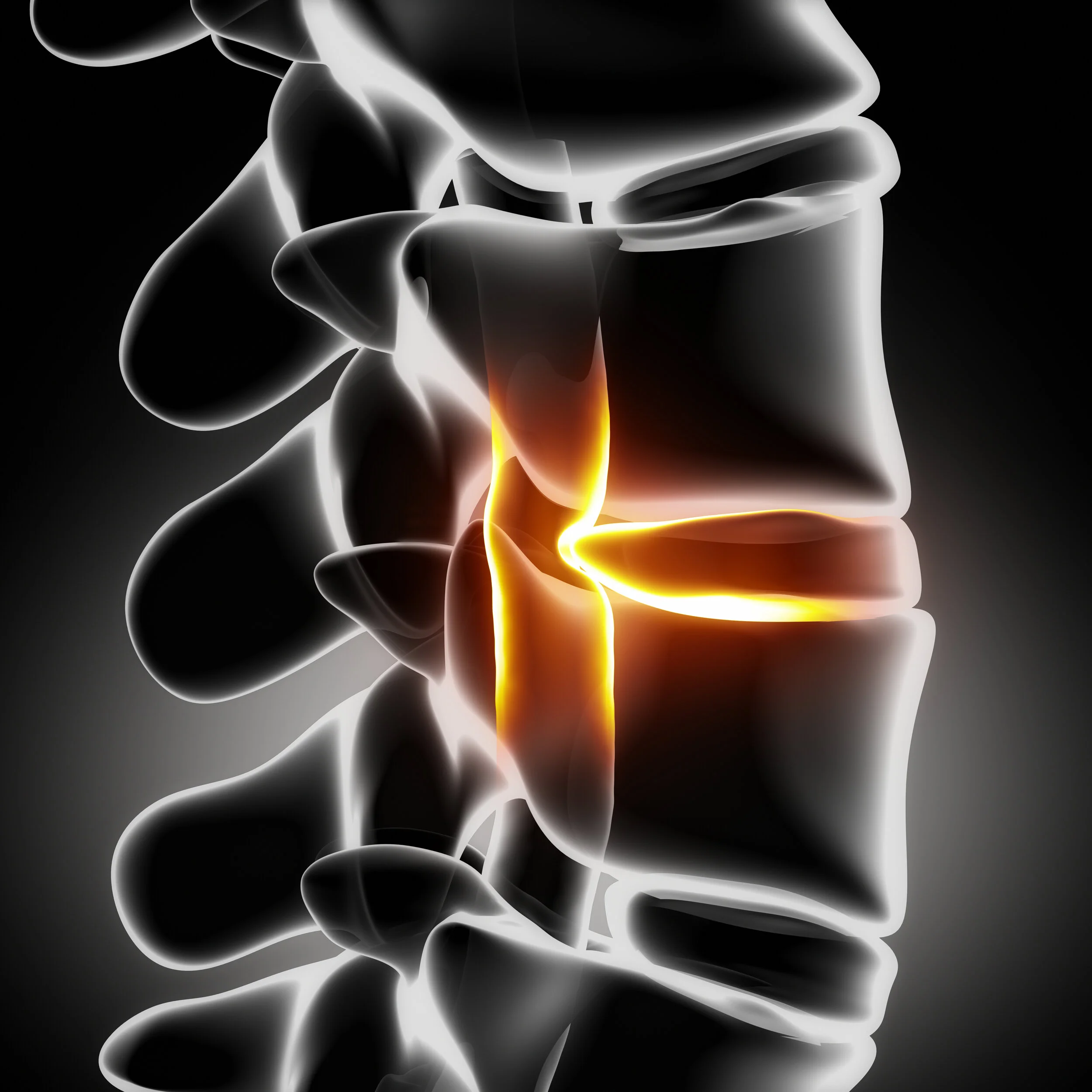
Effects of Vertebral Axial Decompression on Intradiscal Pressure, Journal of Neurosurgery 1994
This study examined the effect of vertebral axial decompression on pressure in the nucleus pulposus of lumbar discs. Intradiscal pressure measurement was performed by connecting a cannula inserted into the patients L4/L5 disc space to a pressure transducer. Changes in intradiscal pressure were recorded at resting state and while controlled tension was applied by the equipment to a pelvic harness. Intradiscal pressure was decreased in the nucleus to below -100 mm HG. Our system also employs a pelvic harness, but with a trapezoid shaped insert affixed to the inside of the harness, placed directly against the lumbar spine. This design feature increases direct friction and localizes decompression force. It follows that our custom lumbar belt would yield a more effective decompression force.
G. Ramos MD, W Martin MD ; Journal of Neurosurgery 1994
Long Term Effect Analysis of IDD Therapy in Low Back Pain: A Retrospective Clinical Pilot Study
Decompression treatment rendered “good” to “excellent” relief in 86% of patients with herniated discs and 75% in patients with facet arthrosis
Traction yielded no “excellent” results in patients with herniated discs and only 50% “good” to “excellent” results with patients who were diagnosed with facet arthrosis.
Norman Shealy, MD, Phd, American Journal of Pain Management, Vol. 7 No. 2, April 1997
Static Traction vs. Decompression vs Dynamic mVAST
Patients treated with traction compared to a control group that had simulated traction demonstrated no significant differences in outcome.
Traditional traction does not produce spinal decompression.
Decompression has been proven as an effective treatment for herniated and degenerative disc disease, by creating a negative intradiscal pressure
Weber H., Traction therapy in sciatica. J Oslo City Hosp. 1973;23(10):167-176
Comparing Static Traction to Motorized Decompression vs. our SoloTrac Platform (mVAST)
Spinal decompression has shown to decompress the disc space, and in the clinical picture of low back pain is distinguishable from conventional spinal traction.
Ramos G, Martin W., Effects of vertebral axial decompression on intradiscal pressure. J Neurosurgery. 1994; 81:350-353
Mangion, et al concluded that any benefit derived from continuous traction devices was due to enforced immobilization rather than actual traction.
Mangion, et al; A controlled trail of continuous lumbar traction in back pain and sciatica. Br J Rheumatol. 1986; 25:181-183
(Note: Our platform does not apply continuous motorized traction and does not enforce immobilization. Our method applies graded decompression force similar to the dynamic unloading/re-loadiong action of a coil spring. Our platform can emulate the force, angle, and amplitude of motorized decompression systems, but with the added comfort and safety afforded by using elastic resistance material. Additionally, our system has the added advantage of a component not used by other systems: We affix a grip pad into our custom lumbar belt. This increases friction between the belt and the lumbar spine, thus yielding a grip that is localized directly at the lumbar spine. Other systems rely on padded columns that press into the pelvic bones, or a lumbar harness that generally grips the pelvis, without direct localized friction against the lumbar spine.
~Matthew Brown, D.C.
Regenerative Potential?
Note: Repeating cycles of unloading and loading will maximize hydraulic-type imbibition. This is where our platform excels compared o motorized systems.
~Matthew Brown, D.C
Disc Distraction Shows Evidence of Regenerative Potential in Degenerated Intervertebral Discs, SPINE 2006
Conclusion:
Disc repair fundamentally depends on the stage of disc degeneration.
This study with respect to previous reports, confirms that disc distraction enhances hydration in the degenerated disc and may improve disc nutrition via the vertebral endplates.
Thorsten Guehring, MD, et al; Department of Orthopedic Surgery, University of Heidelberg, Germany
SPINE Volume 31, Number 15, 2006
Decompression Reduces Chronic Back Pain: 4 Year Study
91% - resumed normal daily activities
87% - working or retired without back pain
71% - had 50% reduction in pain immediately after treatment
86% - showed 50% or better pain reduction at four years.
52% - pain level of zero
Decompression Reduces Chronic Back Pain: 4 Year Study
Summary:
“After 4 years, 52% of respondents reported a pain level of zero. Thus, pain relief not only lasted but improved.”
R. Odell, MD
Excellent indicator that decompression treatment results last.
R. Odell MD, D. Boudreau DO, Anesthesiology News March 2003
Opioid Epidemic? Chiropractic care reduced opioid demand by 49%.
This was verified for Medicare disability beneficiaries. Studies were performed at the Veterans Administration Hospital System.
American Academy of Pain Medicine (AAPM) 2019 Annual Meeting: Abstract 107. Presented March 08, 2019.
Whedon JM, Toler AWJ, Goehl JM, Kazal LA. Association between utilization of chiropractic services for treatment of low-back pain and use of prescription opioids. Journal of Alternative and Complementary Medicine 2018 June 24:6. https://doi.org/10.1089/acm.2017.0131
Weeks WB, Goertz CM. Cross-sectional analysis of per capita supply of doctors of chiropractic and opioid use in younger medicare beneficiaries. Journal of Manipulative & Physiological Therapeutics 2016;39(4):263–6.
Lisi AJ, Corcoran KL, DeRycke EC, Bastian LA, Becker WC, Edmond SN, Goertz CM, Goulet JL, Haskell SG, Higgins DM, Kawecki T, Kerns RD, Mattocks K, Ramsey C, Ruser CB, Brandt CA. Opioid Use Among Veterans of Recent Wars Receiving Veterans Affairs Chiropractic Care. Pain Medicine 2018 Sep 1;19(suppl_1):S54-S60. doi: 10.1093/pm/pny114. PubMed PMID: 30203014.
Nutrients flow in. Waste products flow out. This phenomenon is described thoroughly on our web site. The Human Spinal Disc nucleus can handle low oxygen concentration, but is vulnerable when extra-cellular glucose levels drops too low for a period of several days. The disc needs to imbibe fluid to stay strong, so a drop in proteoglycan content can initiate the downward spiral of disc degeneration within weeks or months. We must keep spine “hydraulics” flowing, especially after a traumatic episode!
Horner H, Urban J P G 2001 The effect of nutrient supply on viability of cells from the nucleus of the intervertebral disc. The International society for the study of the Lumbar Spine, Edinburgh UK”
“Our methodology can emulate the physical decompression force of high tech motorized systems at a fraction of the cost.“
While we cite our own clinical trial results above, there are many attributes about the general topic of “spine decompression” already published in scientific literature that also applies to our platform. Proper application of mVAST method can deliver decompression force, angle, and amplitude almost identical to the algorithms programmed into motorized systems. Any correlation or citation of published studies is intended to consider physiological effects that might be produced with proper application of our platform. When you consider the gratifying comfort and safety our system offers utilizing elastic resistance material, along with the advantage of applying multiple vectors in a single encounter - ours is as effective or better than motorized systems costing 10x more, and the savings can then be passed on to the consumer. When “white coat syndrome” is eliminated, and golgi tendon organ reflexes are desensitized - the encounter becomes exceptionally gratifying for the user.”
~Matthew Brown, D.C.
Using First Principles to Build a Better Product
If you wanted to remove a cork from a wine bottle, which technique is more effective? Gripping and pulling the cork with pliers? Or screwing a corkscrew into the center of the cork and then popping the cork out? When considering different kinds of “spine decompression,” you should consider if the decompression table is designed to press into the pelvic bones with 2 vertical posts . . . vs. concentrating the grip directly against the lumbar spine. We have designed a custom lumbar belt, combined with a triangular shaped pad that is affixed into the belt in order to maximize friction and “grip” directly at the lumbar spine.
-
It all begins with an idea. Dr. Matthew Brown was trained to use high tech decompression machines. However, he realized the design might be more comfortable and more effective by using a gliding platform and elastic resistance material.
-
After searching the world for such a product, he realized the concept had not yet been invented. So he built multiple iterations and got the patent. People loved the therapy - so he kept moving forward.
-
This turns out to be one of the most challenging steps - convincing the industry there is a better way to decompress the spine.

By Mark Studin DC, FASBE(c), DAAPM
Eric Kaplan DC, FIAMA
Reference: Studin M., Kaplan E. (2022) Vertebral Subluxation Complex vs. Patho-Neuro-Biomechanical Lesion, American Chiropractor, 41 (9) Pgs. 18, 20 , 22, 24
Is Chiropractic a philosophy, an art, and a science? Or is it a science, utilizing an art, that recognizes a philosophy? Are you purely a “subluxation-based” practitioner, or only functioning in the pain model and anti-subluxation, or a combination of both? Are you at a chiropractic school that has banned the word subluxation, or a school that mandates the use of that concept? As I pose those questions to chiropractic practitioners, it is too often confusing for them to answer. Imagine the confusion of an unknowing public, or politicians trying to figure out if their support is prudent. Our profession is a “niche’” industry and these disparate beliefs hurt us all no matter your practice model.
Subluxation is not a word that our profession should shy away from. It has been used since the inception of our profession and we have used that to “brand” chiropractic. However, as we have evolved with continual new evidence in the scientific community, we should progress and embrace the changes that will be the core of expanding utilization beyond our (approximate) 9% of the population.
When we look carefully at the vertebral subluxation complex [VSC], Leonard Faye (1966) helped developed that concept. However, there were many others who contributed, and all were considered “theorists” during their time because science had not caught up to those theories. What makes Faye, DD and BJ Palmer and so many others so remarkable are that it was so close to what we know now to be factual, yet it was derived in the absence of today’s technology that has afforded us so many answers.
Faye’s model was congruent with those from previous theorists that chiropractic vertebral subluxation (CVS) may lead to pathophysiology and then pathology. He hypothesized that normal physiological processes would be restored, and the “life forces” would be unblocked by correcting the CVS. He proposed that the objective was to develop an examination rationale to look at the locomotor system as a whole with the spine as “part of a closed kinematic system. The rationale for adjusting included finding the fixation, mobilizing the fixation, and rechecking to confirm improvement. Faye in conservation discussed creating the concept of CVS as “a complex clinical entity” in 1963 as comprising pathophysiological changes associated with “one or more of the following: Neuropathophysiology Kinesiopathology, Myopathology, Histopathology, and Biochemical Pathology.
If it wasn’t for Faye and others, who piggybacked and expanded on DD and BJ’s “vitalistic” theories, where would science have looked? Most scientific breakthroughs were founded on theory, then tested to figure out why in the validation process in the proverbial “laboratory.” Chiropractic was and is no different. Here, in part is what science has explained about chiropractic, subluxation, and biomechanics.
Evans (2002) reported “…on flexion of the lumbar spine, the inferior articular process of zygapophyseal joint moves upward, taking a meniscoid with it. On attempted extension, the inferior articular process returns toward its neutral position, but instead of re-entering the joint cavity, the meniscoid impacts against the edge of the articular cartilage and buckles, forming a space-occupying "lesion" under the capsule: a meniscoid entrapment. A large number of type III and type IV nerve fibers (nociceptors) have been observed within capsules of zygapophyseal joints. Pain occurs as distension of the joint capsule provides a sufficient stimulus for these nociceptors to depolarize. Muscle spasm would then occur to prevent the impaction of the meniscoid. The patient would tend to be more comfortable with the spine maintained in a flexed position because this will disengage the meniscoid. Extension would therefore tend to be inhibited. This condition has also been termed a "joint lock" or "facet-lock" the latter of which indicates the involvement of the zygapophyseal joint.” P. 252-253
NOTE: The Evans article has been cited 265 times, as recently as numerous times in 2022. It has been reported that anything over 25 times is considered impactful in the scientometric indicator.
The dislodged meniscoid renders biomechanical pathology or aberrant loading of the spinal column. Panwar and Hamza (2022) reported spinal inability to carry spinal loads…includes the clinical consequences of neurological deficits and/or pain.” P. 53
Panjabi (2006) described the negative sequella of pathobiomechanics in a stabilization-destabilization scenario involving corrupted neurological transducers. As Evans reported on the meniscoid entrapment, the process includes distention and firing of the joint capsule. The joint capsule as reported by Solomonow (2009) is comprised of ligaments for both mechanical and sensory functions. Dougherty (2020) reported Pacinian (crimp receptors) and Ruffini (stretch receptors) Corpuscles, Golgi ligament organs, and free nerve endings (nociceptors) in the joint capsule. These provide proprioception and mechanoreception afferently and are all considered somatosensory receptors.
As a result of the biomechanical instability, as reported by Solomonow (2009) there is a compensatory ligamento-muscular reflex thatmay be inhibitory or excitatory, as may be fit to preserve joint stability; inhibiting muscles that destabilize the joint or increased antagonist co-activation to stabilize the joint. Panjabi (2006) then reported “The corrupted muscle response pattern leads to corrupted feedback to the control unit via tendon organs of muscles and injured mechanoreceptors [and nociceptors], further corrupting the muscle response pattern. (p. 669)
Sampath et. Al (2017) reported “A key causative mechanism responsible for the perception of pain is nociception that occurs at the site of tissue injury… These biochemical markers include various neuropeptides such as neurotensin, oxytocin, substance-P (SP), and orexin-A… These chemicals are primarily released at the injury site, they also result in the initiation of an inflammatory process which further results in the production of numerous pro-inflammatory and immuno-regulatory cytokines and neurotransmitters.” p. 120
The meniscoid entrapment as described by Evans (2002) and the type III and IV nociceptor reaction at the zygapophyseal joints explain where there is bone on the nerve and aberrant neurological sequella. Cramer et. Al (2002) reported that a chiropractic high velocity, low amplitude adjustment creates joint gapping and normalizing of spinal biomechanics. These are topics discussed in other papers but underscore that biomechanics can be pathological and create negative neurological sequelae.
The result of biomechanical failure is remodeling, based upon Wolff’s Law. Wang et. Al (2019) reported “According to Wolff’s law, bones in the living body will adapt to mechanical loads under which they are placed. If loads on a particular bone increase, the bone will remodel to become thicker and stronger to resist the loads. The inverse is also true; if loads on a bone decrease, the bone will become thinner and weaker. Then, does the morphology of human bones continue to change under long-term strains after skeletal maturity? The answer is also YES. Based on clinical observations and a series of scientific studies, the dynamic deformation of human bones continues under long-term strains even after skeletal maturity.” P. 2636 The same responses occur in connective tissues according to Davis Law which is a corollary of Wolf's Law.
The above articles represent 20 years of evidence in the literature and is rending answers to past theories. Faye’s theory of the 5 components of the VSC also validates DD and BJ’s theory of bone on the nerve but now allows for an accurate anatomical location of what bone on what nerve(s). Now that we have an evidence-based affirmation, how do we best position our communication, bill with carriers, and argue in the medical-legal-insurance communities from a legally defensible posture? The answer must be devoid of philosophy, prejudice, and dogma from within the chiropractic profession to prevent furthering the chasm based upon the dichotomy of beliefs.
Although it would be most appropriate to use the term Patho-Neuro-Biomechanical Lesion based on contemporary literature, it would create more confusion with a new description of what chiropractic treats and does not solve the medical-legal-insurance-internal philosophical challenges. Although ICD-10 has added vertebral subluxation complex, it is not directed at chiropractic. Gwilliam (2012) reported, “According to General Equivalency Mapping (GEMs), the commonly used ICD-9-CM code of 739.1 (non-allopathic lesions; cervical region cervicothoracic region) may be replaced with M99.01, which is “segmental and somatic dysfunction of the cervical region.” This differs little from ICD-9-CM and still does not use the word “subluxation.” However, nearby we find the code M99.11, which is defined as “subluxation complex (vertebral) of the cervical region.” This sounds just like the verbiage most chiropractors use, but GEMs point to this code back to 839.00, not 739.1 in ICD-9-CM. This is the code for “closed dislocation, cervical vertebra, unspecified,” which implies that the definition is still not geared towards the chiropractic model.”
The answer has been revealed in the ICD-10 code in the M99.8X code set:
M99.81 Other biomechanical lesions Cervical region
M99.82 Other biomechanical lesions Thoracic region
M99.83 Other biomechanical lesions Lumbar region
M99.84 Other biomechanical lesions Sacral region
M99.85 Other biomechanical lesions Pelvic region
Once you establish a biomechanical lesion, you can further define the pathological sequella in your patient with additional diagnostic codes ranging from muscular and neurological to systemic or any other clinically valid findings you conclude on your patient. The M99.8X diagnostic codes best reflect the evidence in the literature giving a causative foundation for the 5 components of the VSC and offering a pathway to position the chiropractic profession as leaders in the diagnosis and treatment of mechanical spine pathology. It is evidence-based and consistent with the medical, legal, and insurance industries.
All our schools need to teach the 5 components of the VSC, and the evidence to support the conclusion as that will help define who we are. All our political organizations need to put aside philosophical differences, take blinders off, and evolve in accordance with the evidence in the literature. However, all academic and political entities should rally around the diagnostic conclusion of biomechanical lesions as that is consistent with the evidence in the literature and the insurance industry. It allows us to mainstream our conclusions and avoid confusion in the healthcare industry. It is the common bond for every DC and allows each practitioner to further define care based upon additional diagnosis, how they choose to practice within their lawful scope.
REFERENCES
Beliveau, P. J. H., et al. "The chiropractic profession: a scoping review of utilization rates, reasons for seeking care, patient profiles, and care provided. Chiropr Man Therap. 2017; 25: 35.
Senzon, Simon A. "The chiropractic vertebral subluxation part 9: complexes, models, and consensus from 1979 to 1995." Journal of Chiropractic Humanities 25 (2018): 130-145.
Evans, David W. "Mechanisms and effects of spinal high-velocity, low-amplitude thrust manipulation: previous theories." Journal of manipulative and physiological therapeutics 25.4 (2002): 251-262.
Plomp, Reinier. "The significance of the number of highly cited papers as an indicator of scientific prolificacy." Scientometrics 19.3 (1990): 185-197.
Panwar, Yudhisthir, and Yusuf Gambo Hamza. "Clinical instability spine and lower back pain." International Journal of Innovative Research in Science Engineering and Technology, (2022): 53-66.
Panjabi, M. M. (2006). A hypothesis of chronic back pain: Ligament subfailure injuries lead to muscle control dysfunction. European Spine Journal,15(5), 668-676.
Solomonow, M. (2009). Ligaments: A source of musculoskeletal disorders. Journal of Bodywork and Movement Therapies,13(2), 136-154
Dougherty, P. (2020). Chapter 2: Somatosensory systems. Neuroscience Online. Retrieved from http://neuroscience.uth.tmc.edu/s2/chapter02.html
Kovanur-Sampath, Kesava, et al. "Changes in biochemical markers following spinal manipulation-a systematic review and meta-analysis." Musculoskeletal Science and Practice 29 (2017): 120-131.
Wang, Juan, et al. "Law of dynamic deformation of bone." Chinese Medical Journal 132.21 (2019): 2636-2637.
Gwilliam (2012), Chiropractic’s Favorite Word Finds its place in ICD-10… Or Has it? Retrieved from: https://icd10monitor.com/chiropractic-s-favorite-word-finds-its-place-in-icd-10-cm-or-has-it/